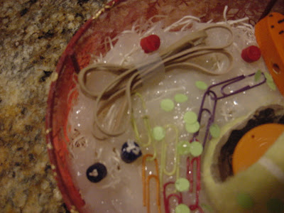
Here is a model of a functioning animal cell using simple household, office and construction supplies. Each product was handpicked for its resemblance to portrayal of organelles in in various cell illustrations and models. Here is a list of organelles. The numbers correspond to the numbers next to each item in the photo to the right. 1. Cell Membrane (wire and cloth basket) 2. cytoplasm (window sealer/packing straw) 3. Nucleus (tennis ball) nuclear pores (black spots on ball) 4. nucleolus, chromatin (orange cap, black space in ball) 5. mitochondria (the one represented here is different from the one I ended up using, which is a wooden carrot) 6. one of three chromosome pairs used in sub model (metal braces)7. ribosomes (hole-punched paper) 8. Golgi
 apparatus (taped rubber bands) 9. Endoplasmic reticulum (linked paper clips) 10. vesicles (red crayon. Lysosomes will later be shown as blue crayons with straw at center to represent digesting bacteria.) The flagellum will be represented as a cut hair tie.
Here is the nucleus inside the cell.Its function is the production of DNA The membrane is the wire a
 nd mesh basket and the straw and sealer(clear goo) is the cytoplasm. The yellow outer of the nucleus is the nuclear envelope. The shiny orange cap is the nucleolus and the black in represents the chromatin. The black spots on the outside of the tennis ball (or shall I say nucleus) are nuclear pores through which ribosomes and mRNA enter and exit the nucleus.
The paper clips, here, represent the ER (endoplasmic reticulum.) Its function is to put proteins to use. This is also where proteins fold. Glucose and other products are made there.The green dots are ribosomes. They are key to amino acid production. When found on the ER that ER is called rough ER. The ER lacking ribosomes is the
 smooth ER.
The orange ellipse in the photo below is the mitochondria. This is where ATP (fuel of cellular energy) is produced. The fused bands is the
 Golgi apparatus. These organelles receive vesicles with particles for construction of important molecules and sends vesicles out to secrete waste. The vesicles are the red cylinders. When they collect materials from outside the cell, it is called endocytosis. When they excrete materials it is called ectocytosis.
The blue cylinders are lysosomes. They digest bacteria (little straw in cylinder) to use particles or secrete them.
The "tail" here is the flagellum. Its function is cellular movement. S
 ome cells have flagella. Others use cilia for movement and other functions.
Here is the complete, working cell. Now Let's take a look at what goes on in DNA production.

This is a paired chromosome ( the two metal braces) in the nucleus. Each human cell has 46 chromosomes. When a cell is ready to reproduce it copies each chromosome. They are then paired, like so. The colored wires represent the DNA that chromosomes are made up of. DNA is twisted in a "double helix" pattern. The rungs are genetic code. They are made up of four base units. When DNA is reproduced it, first, splits as shown above. Each side, then connects with a copy that has been made.

When The base units for DNA (adenine, guanine, cytosine and thymine) and other DNA products are needed mRNAs (messengers) take the information to ribosomes. There the information is read and tRNAs (transmitters) are put to use to make the necessary materials. The red spiral is an mRNA being read by the ribosome (green dot) the orange spirals are tRNA entering and leaving the ribosome.
It took hours to put together a model of the microscopic cell. It doesn't, of course, function as a live cell does. Cells are amazingly intricate. In the time it took me to make this model, millions of cells were born in my body. Millions of cells went about their usual business to keep me functioning normally. Millions of cells carried out tasks with such complicated instructions that it would make a city planner's head spin. There is, perhaps, much we can learn from the little cell. It functions smoothly. It works efficiently. It works together with other cells in great harmony. It identifies problems and takes care of them promptly. When a problem does arise, however, the results can be disastrous (cancer, disease.) If we might learn to take the efficiency and learn from the mistake of "bad cell multiplication" who knows what we might accomplish.
PS: for more information and photos on mitosis contact me at hexinduction@yahoo.com
Post Post Script.: It turns out today (2/17) I was able to use my cell model to teach a friend's young children about cells. They were captivated by the colors. NOTE: teach kids with COLORS!!! haha.


No comments:
Post a Comment